
Colorectal cancer is a common malignancy arising from the colon or rectum and is one of the important global health concerns. Most colorectal cancers develop from the glandular lining of the bowel and are classified as adenocarcinomas. The disease may initially develop from precancerous polyps and can progress gradually through genetic and cellular changes.
Management of colo-rectal cancer depends on the tumor’s location, stage, histological characteristics, molecular profile, and the patient’s overall health, and can involve a combination of surgery, radiotherapy, chemotherapy, targeted therapy, immunotherapy, and Ayurvedic treatment modalities. Advances in screening, imaging, surgical techniques, systemic therapies, and multidisciplinary management have significantly improved the ability to diagnose and treat colorectal cancer and have contributed to better outcomes for many patients.
Case Overview
A female patient in her late 40s was diagnosed in July 2025 with moderately differentiated adenocarcinoma of the low rectum. The patient underwent radiotherapy and systemic chemotherapy, along with Integrative Ayurvedic Treatment under the care of Dr. Ravi Gupta.
Dr. Ravi Gupta, M.D. (Ayurveda), is an Ayurveda Cancer Consultant specializing in integrative Ayurvedic oncology and traditional cancer care. With extensive clinical experience in Ayurveda and a focused approach to cancer care, he provides individualized treatment plans incorporating Ayurvedic medicines, diet and lifestyle guidance, Rasayana therapy, and Panchakarma.
Initial Diagnosis
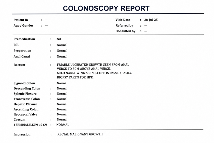
The patient underwent colonoscopy on 28th July 2025. The examination revealed a variable ulcerated growth extending from the anal verge to approximately 5 cm above the anal verge, associated with mild narrowing. The scope was passed easily through the narrowed segment. The remainder of the colon and terminal ileum was reported as normal.
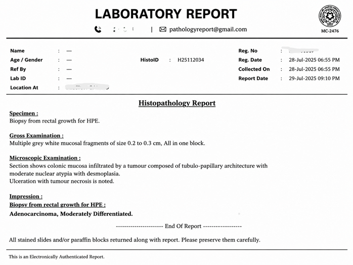
Biopsy of the rectal growth established the diagnosis of moderately differentiated adenocarcinoma. Histopathology demonstrated colonic mucosa infiltrated by a tubulo-papillary tumor with moderate nuclear atypia and desmoplasia. Ulceration with tumor necrosis was also noted. The oncology documentation subsequently described the diagnosis as Grade II, moderately differentiated rectal adenocarcinoma.
Pelvic MRI dated 2nd August 2025 further characterized the primary tumor. It was a low-lying rectal tumor, with its lower extent approximately 20 mm from the anal verge and below the anterior peritoneal reflection. The tumor had an approximately 56-mm craniocaudal extent and a circumferential configuration.
The lesion involved the lower rectum and anal canal, including the internal sphincter and intersphincteric region. A perianal fistulous tract was described at approximately the 11 o’clock position. The lesion appeared to involve or be adherent to the mesorectal fascia and adjacent posterior vaginal wall, with involvement of the right levator ani region described on the MRI. Multiple heterogeneous mesorectal lymph nodes were identified, with the largest measuring approximately 8 × 5 mm. Heterogeneous left internal iliac lymph nodes were also present, with the largest measuring approximately 9 × 5 mm.
These findings represented a locally advanced low rectal malignancy with significant local anatomical involvement and suspicious regional nodal disease.
Multimodality Treatment
She received 28 cycles/fractions of radiotherapy which she completed by September 2025, together with systemic chemotherapy (CAPOX/SOX) and subsequent consolidation treatment.
In addition to her current oncology treatment, the patient also received Integrative Ayurvedic Treatment from Dr. Ravi Gupta. The integrative Ayurvedic approach was intended to improve the patient’s overall wellbeing and successful cancer-treatment journey.
MRI Assessment After Treatment
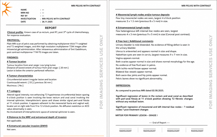
The pelvic MRI dated 26th November 2025 demonstrated significant regression of the rectal and anal canal lesion compared with the pretreatment MRI. The previously extensive lesion had markedly decreased and showed predominantly T2-hypointense characteristics compatible with treatment-related fibrosis. Importantly, the MRI did not demonstrate diffusion restriction or an abnormal ADC pattern suggesting obvious viable residual tumor.
The previously described perianal fistulous region showed T2 fibrotic changes without an obvious residual lesion. There was also significant regression of the previously described mesorectal and left internal iliac lymph nodes.
The radiologist assigned MR Tumor Regression Grade I (MRTRG Grade I) to the primary lesion. This represents a highly favourable radiological treatment response, with the imaging appearance predominantly reflecting fibrosis rather than a definite residual tumor mass.
PET-CT Demonstrating Metabolic Response
A whole-body PET-CT performed on 23rd February 2026 provided additional evidence of the favourable response.
At the treated rectal bed, only minimal non-FDG-avid soft-tissue thickening remained at the anorectal junction and upper anal canal. The maximum wall thickness was approximately 6.5 mm. The PET-CT described this appearance as a burnt-out lesion with post-radiotherapy changes.
No obvious hypermetabolic residual mass was identified within the rectum or anal canal. The mesorectal space and mesorectal fascia appeared unremarkable. There was also no active FDG-avid perianal fistula or collection in the ischioanal or ischiorectal fossae.
The previously noted lower mesorectal node was now tiny and non-FDG avid and was described as burnt out. Other small pelvic and retroperitoneal lymph nodes were also non-FDG avid and considered reactive.
Most importantly, the PET-CT demonstrated no evident FDG-avid distant metastatic disease. There was no evidence of FDG-avid peritoneal carcinomatosis or other distant hypermetabolic malignant lesions.
Follow-Up MRI
The subsequent MRI performed on 4 May 2026 continued to show a favourable treatment response. Only mild eccentric circumferential T2-hypointense wall thickening remained at the anorectal junction and upper anal canal. The maximum wall thickness was approximately 8 mm, while the involved segment measured approximately 37 mm, compared with approximately 56 mm at baseline.
The lower margin of the residual soft-tissue thickening was approximately 27 mm from the anal verge. Minimal surrounding fibrotic changes were present in the anterior mesorectal space. The report described focal adherence to the adjacent mesorectal fascia and the right half of the posterior vaginal wall.
Importantly, the radiological impression characterized the abnormality as: “Burnt-out wall thickening with post-RT changes.” No abnormal mesorectal lymph nodes were identified at this examination.
Overall Treatment Response
The serial investigations demonstrate a major radiological response of the locally advanced low rectal adenocarcinoma following multimodality treatment.
At diagnosis, the patient had a low-lying circumferential rectal tumor extending into the anal canal, with involvement of important local anatomical structures and suspicious mesorectal and pelvic lymph nodes.
Following radiotherapy, and systemic chemotherapy, and Integrative Ayurvedic Treatment under Dr. Ravi Gupta, there was substantial regression of the primary lesion and nodal abnormalities. Subsequent imaging demonstrated predominantly fibrotic tissue, absence of significant FDG uptake, and no evidence of distant metastatic disease.
Conclusion
This case represents a locally advanced low rectal adenocarcinoma showing marked response following multimodality cancer treatment, with serial MRI and PET-CT demonstrating substantial regression of the primary lesion and previously suspicious lymph nodes.
The most recent available imaging shows predominantly post-radiotherapy fibrotic/burnt-out changes, no abnormal mesorectal lymph nodes, and no evident distant metastatic disease.
The patient’s treatment journey also included Integrative Ayurvedic Treatment under the care of Dr. Ravi Gupta, which contributed to the overall management of the condition and was associated with a favourable response to treatment for rectal cancer.
Contact Dr. Ravi Gupta – National & International Patients
Patients from across India and overseas can contact Dr. Ravi Gupta, M.D. (Ayurveda), Ayurveda Cancer Consultant, for personalized consultation and integrative Ayurvedic cancer care. Online consultations are available for international patients who wish to discuss their diagnosis, treatment history, medical reports, and supportive Ayurvedic care from their home country.
Contact Dr. Ravi Gupta:
📞 +91-9819274611
🌐 www.cancerinayurveda.com Patients are encouraged to share their latest medical reports and treatment details while requesting a consultation.